Go to page content

Magnetic resonance imaging (MRI) examination

MRI examination provides detailed images of the body without the use of radiation. The examination utilizes a strong magnetic field and radio waves to visualize water in various forms within the body.

MRI examination is a diagnostic imaging method used to examine different parts and tissues of the body. The abbreviation for magnetic resonance imaging is MRI. 

Watch the video: Magnetic resonance imaging. The video is produced by HUS and includes subtitles in Finnish, Swedish, and English. 

You will receive patient instructions detailing how to prepare for the examination. Some MRI examinations require no preparation, while others may require you to fast, for example. 

Reserve plenty of time for your visit. The examination typically lasts 30–60 minutes. Patients requiring urgent imaging may be examined ahead of you. 

Remember to fill out the pre-information form and bring it with you to the examination. A radiographer will review the form with you before the examination. 

If you have metal or metallic parts in your body, contact the MRI unit in advance. For example, a pacemaker, prosthesis, intracranial surgical clip, shrapnel, or medication pump may require special planning and preparation or may prevent MRI examination. 

Joint prostheses or metal implants fixed to bone during orthopedic surgery usually do not prevent imaging, but inform the MRI unit staff about them. Dental fillings and similar materials fixed in the mouth are generally safe. 

If you know or suspect that you are pregnant, inform your treating physician or the MRI unit staff at the latest when you arrive for the examination. MRI can be performed at any stage of pregnancy if medically necessary. However, MRI is generally avoided during early pregnancy as a precaution. The decision to perform MRI on a pregnant patient is made by a physician. 

The use of MRI contrast agents is not recommended during pregnancy, but may be considered if medically necessary. The decision to use contrast agents is made by a physician. Contrast agents may be administered to breastfeeding patients. 

During the MRI examination, you lie on a padded examination table. The table is moved into the scanner so that the area being imaged is positioned in the center of the scanner. The opening of the scanner is approximately 70 cm in diameter and is open at both ends, well-lit, and ventilated. You will enter the scanner either head-first or feet-first. You will be given a call button to hold in your hand to contact the radiographer if needed. 

The radiographer operates the MRI scanner from a computer in an adjacent room, behind a window. The radiographer can see you throughout the imaging. You can communicate with the radiographer via microphone if necessary. 

The scanner produces loud tapping or buzzing sounds during the imaging. You will be provided with hearing protection and possibly earplugs. You can listen to music through the hearing protection. The changing magnetic field may also cause slight vibrations in the examination table. It is very important to remain still and relaxed during the imaging. 

You may be given an MRI contrast agent intravenously during the imaging. The physician determines the dosage. The contrast agent helps in interpreting the images. MRI contrast agents contain gadolinium (not iodine) and are excreted from the body via urine within a few hours. 

Allergic reactions to MRI contrast agents are rare. You can ask your referring physician or the MRI unit staff for more information. 

MRI examination typically last 20–30 minutes. A comprehensive MRI may take over an hour. 

About 10% of patients experience claustrophobia. Inform the MRI unit if you suffer from claustrophobia and arrive early for your appointment. Familiarizing yourself with the scanner and, if necessary, receiving a sedative premedication can help the examinatio proceed smoothly. Contact your treating unit well in advance regarding possible premedication. You may bring a support person with you. The same safety rules apply to support persons as to patients. 

MRI does not require follow-up and does not impose restrictions on normal activities. Any contrast agent used during the examination is excreted via urine within a few hours. 

Breastfeeding mothers must take a break from breastfeeding after the examination. Milk expressed during the break should be discarded. The healthcare staff will provide detailed instructions to breastfeeding mothers after the examination. 

A radiologist will review and interpret the MRI images and write a report. The physician who referred you for the examination will inform you of the results and discuss further treatment. Contact your care unit if you do not have a scheduled follow-up appointment or phone consultation. 

MRI is painless and has not been shown to have adverse effects. However, MRI is not suitable for everyone; certain implants and foreign objects in the body, such as pacemakers or metal fragments, may prevent imaging or require special arrangements. 

If you know or suspect that you are pregnant, inform your treating physician or the MRI unit staff at the latest when you arrive for the examination. 

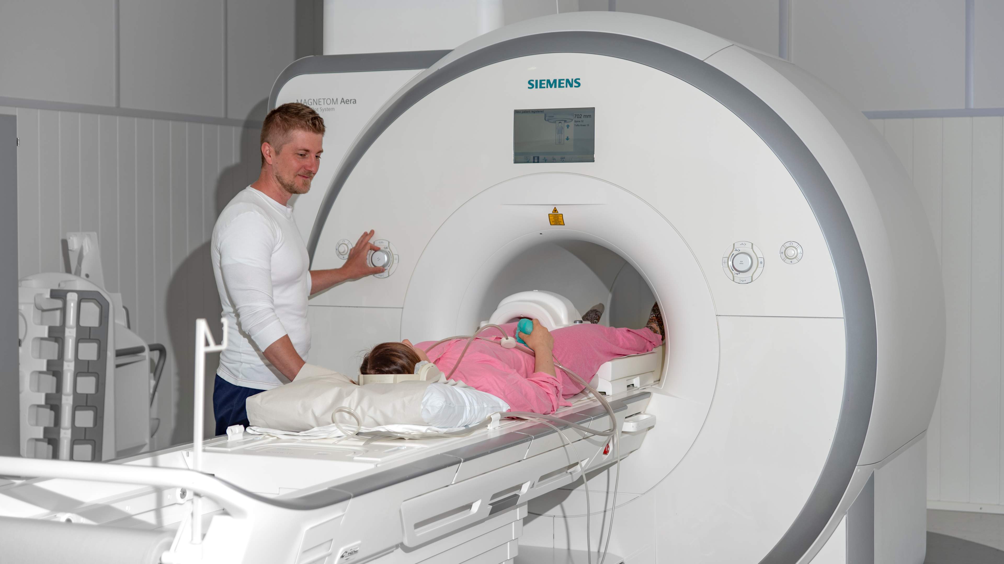
MRI does not use radiation and is a safe imaging method for children, adolescents, and pregnant individuals. The operation of the MRI scanner and image formation are based on the use of magnetic fields and radio waves. Imaging coils are also used to collect data from different parts of the body. MRI is particularly suitable for examining the head, spine, muscles, blood vessels, and internal organs. The duration of an MRI examination ranges from 20 minutes to over an hour, depending on the area being imaged. 

In Finland, MRI scanners commonly used for patients have a magnetic field strength of 1.5 Tesla or 3 Tesla. The Tesla unit measures magnetic force, with a 1.5 Tesla scanner being approximately 30,000 times stronger than Earth’s gravitational pull. It is important to note that MRI scanners are always on and cannot be turned off between examinations. Therefore, metallic or magnetically sensitive items must not be brought into the MRI room. These include keys, metal-framed glasses, pens, piercings, jewelry, hairpins, scissors, tools, etc. The magnetic field can also damage electronic devices, smartwatches, phones, and magnetic stripe cards such as bank cards. These items must be left outside the MRI room in a locked locker or cabinet. 

Inform the healthcare staff upon arrival if you have any implanted device (i.e. pacemaker, cardiac monitor, neurostimulator, hearing aid, pain pump) or any metallic fragment (i.e. shrapnel or pellet) in your body. These may not necessarily prevent the examination but may require preparation to ensure successful imaging. Following instructions is crucial for safety. The same safety guidelines apply to everyone in the room, including staff and support persons. 

If you are using a medication or nicotine patch, remove it before the MRI examination. The examination may raise body temperature, which could cause the active substance in the patch to be absorbed unnecessarily. Do not wear heavy makeup on the face (some makeup products may contain metallic particles), magnetic false eyelashes, or eyelash jewelry. The MRI room is well ventilated, so it is advisable to wear long-sleeved and long-legged clothing. Ensure that your clothing does not contain metal. You may also be provided with hospital clothing such as pajamas at the imaging site. 

Updated 12.11.2025