Go to page content

Ultrasound examination

An ultrasound examination, also known as echocardiography, is a widely used imaging method.

Ultrasound examination is based on the reflection of sound waves at the interfaces of different tissues and their attenuation depending on the acoustic properties of the tissues. Ultrasound guidance is also used in various procedures, such as collecting tissue and cell samples. 

Ultrasound can be used to examine, among other things: 

  • the abdominal area (gallbladder, liver, pancreas, kidneys, spleen, bladder) 
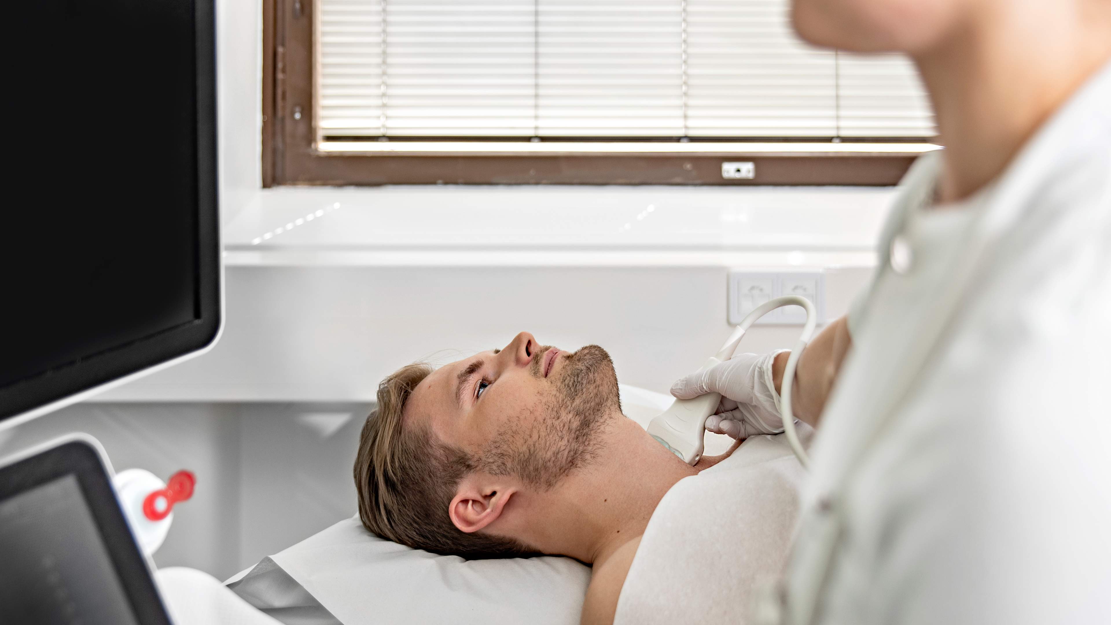
  • the neck area 

  • blood vessels and blood flow 

  • muscles, tendons, or joints 

Watch the video: Ultrasound examination. The video is produced by HUS and includes subtitles in Finnish, Swedish and English. 

Check the patient instructions you received how to prepare for the examination. For example, for upper abdominal ultrasound examinations, you should refrain from eating and drinking beforehand. For lower abdominal examinations, the bladder should be full. 

Ultrasound is a painless and safe examination method. Gel is applied to the skin, and the area is examined by moving the ultrasound probe over the target area. 

In follow-up ultrasound examinations, contrast agents may be administered via a cannula, usually into a vein in the elbow crease. These examinations are performed in a hospital. Minor procedures may also be performed during the ultrasound, such as removing excess fluid with a needle from a joint, abscess, or pleural cavity. Additionally, ultrasound can be used to collect samples from various organs or the mammary gland. Local anesthetic may be applied to the skin and surrounding tissue as needed during procedures. 

The examination typically lasts about 10–30 minutes depending on the area being examined and any procedures performed. 

Ultrasound examination does not require follow-up and does not impose restrictions on normal activities. If a procedure requiring aftercare was performed during the examination, you will receive aftercare instructions. 

The physician who referred you for the examination will inform you of the results and how treatment will proceed. Contact your care unit if you do not have a scheduled appointment or call time. 

Ultrasound examination is safe and can be performed on children and pregnant women. If a contrast agent is used, there is a small risk of an allergic reaction. Ultrasound does not use X-rays, so there is no radiation exposure to the patient. 

When samples are taken, there is a small risk of infection or internal bleeding, but this is rare. You will receive aftercare instructions when samples are taken, and following them will minimize risks. 

Updated 14.11.2025