
Occasional adverse effects
Occasional adverse effects result from random changes in individual cells. Cellular changes occur naturally in tissues, and the body is capable of repairing them.
The most significant occasional adverse effect associated with radiological examinations or procedures involving radiation is a small risk of cancer. Radiological examinations contribute a very slight statistical increase to the overall incidence of cancer.
In Finland, the risk of dying from cancer due to causes other than radiation is 1 in 5 (one people of five) i.e., approximately 20% of the population will die from cancer at some point in their lives.
Direct adverse effects
Direct adverse effects refer to tissue damage caused directly by radiation. At low radiation doses, no tissue damage occurs—for example, no imaging examinations involving radiation cause direct adverse effects.
However, demanding and prolonged radiation procedures may occasionally result in effects such as skin redness. These rare effects are told separately.
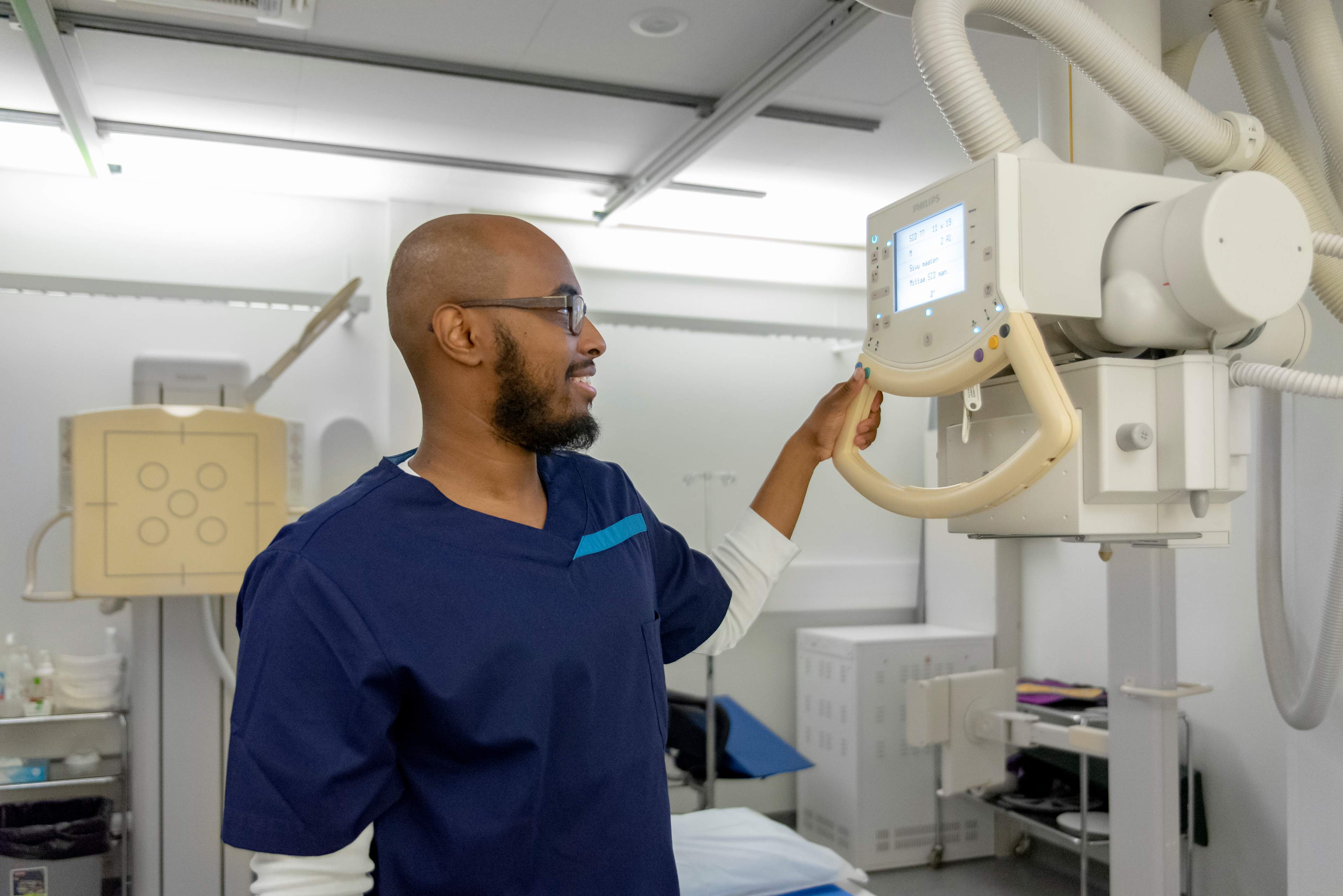
If you know or suspect that you are pregnant and are scheduled for an imaging examination involving radiation, and you have concerns about radiation exposure, please consult the referring physician or the imaging unit's healthcare staff. You can find more information in the section Pregnancy and examinations in the Examinationhub.
Watch the video: Radiation Safety ABC – Can radiation from imaging examinations be harmful? Produced by OYS, with subtitles in Finnish and Swedish.
Radiation doses and estimated risk by examination type
The additional cancer risk depends on the age of the individual. Radiation exposure during childhood results in a higher additional risk than the same dose in adulthood. For a small child, the risk is approximately three times higher, while for a 90-year-old, it is about one-third of the risk compared to a 30-year-old. However, the additional risk for children remains very low.
There are also individual differences in radiation sensitivity, and women are more sensitive to radiation effects than men. Additionally, the sensitivity to radiation varies between different tissues and organs.
The table below provides information on various examinations, their radiation doses, and the estimated probability of radiation-induced cancer risk. The potential cancer risk from a radiological examination depends on the type of examination, the radiation dose, and the characteristics of the individual. It is not possible to precisely estimate the cancer risk for an individual patient.
Table of radiation doses:
Examination | Effective radiation dose (mSv)1 | Equivalent exposure time from all radiation sources 2 | Additional cancer mortality risk (Verbal) | Additional cancer mortality risk (Probability)3 |
|---|---|---|---|---|
Limb X-ray | <0,02 | less than 2 days | almost nonexistent | <1:1 000 000 |
Chest X-ray | 0,02 - 0,2 | 2–12 days | minimal | 1:1 000 000 - 1:100 000 |
Pelvic X-ray, Head CT | 0,2 - 2,0 | 12 days – 4 months | very low | 1:100 000 - 1:10 000 |
Abdominal CT, coronary angioplasty, common angiographies, bone cintigraphy | 2,0 - 20,0 | 4 months – 3 years 5 months | low | 1:10 000 - 1:1000 |
Clarifications for terms used in the table:
1 Effective dose: A quantity that describes the overall health impact caused by ionizing radiation, taking into account radiation-sensitive organs.
2 Average radiation exposure in Finland: Based on 2018 data from the Radiation and Nuclear Safety Authority (STUK), the average annual exposure is 5.9 mSv.
3 Probability: The figures represent statistical additional risk. The exact risk for an individual patient cannot be determined.
Comparison: A four-hour flight at an altitude of 10 km results in a radiation dose of 0.02 mSv.