
The primary indication for small intestine MRI is Crohn’s disease, which often requires repeated imaging in young adults and even children. MRI can also be used to examine the cause of recurrent obstructive symptoms and to detect tumors in the small intestine. MRI is a diagnostic imaging method based on the magnetic properties of the body and does not use X-ray radiation.
Magnetic Resonance Imaging (MRI) of the small intestine
MRI provides detailed anatomical and partially functional images of the small intestine.
You will receive patient instructions detailing how to prepare for the examination.
Remember to fill out the pre-information form and bring it with you to the appointment. A radiographer will review the form with you before the examination.
If you have metal or metallic parts in your body, contact the MRI unit in advance. For example, a pacemaker, prosthesis, intracranial surgical clip, metal fragments, or medication pump may require special planning and preparation or may prevent MRI altogether.
Joint prostheses or metal implants fixed to bone during orthopedic surgery usually do not prevent imaging, but inform the MRI unit staff about them. Dental fillings and similar materials fixed in the mouth are generally safe.
If you know or suspect that you are pregnant, inform your treating physician or at the latest when arriving for the examination. MRI can be performed at any stage of pregnancy if the condition of the pregnant person requires it. As a precaution, MRI is generally avoided during early pregnancy. The decision to perform MRI on a pregnant person is made by a physician.
The use of MRI contrast agent is not recommended during pregnancy, but may be considered if necessary due to the pregnant person’s condition. The decision to use contrast agent is made by a physician. Contrast agent can be administered to a person who is breastfeeding.
Reserve approximately 3 hours for the examination visit.
About 45 minutes before imaging, you will be given a solution containing sorbitol to drink. It is recommended to move around during drinking to help the solution progress through the intestines.
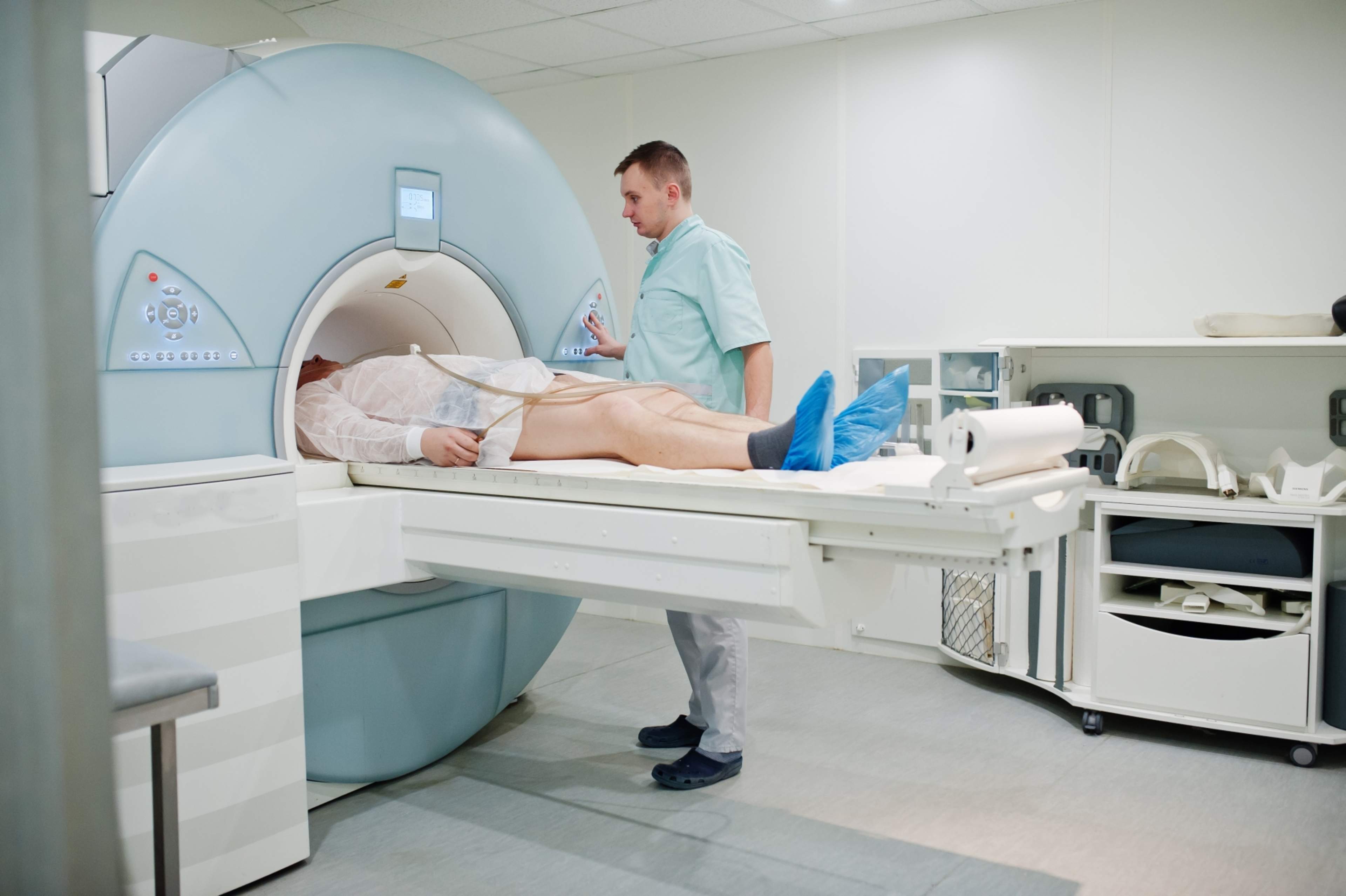
For the imaging, you will be placed on the examination table inside a tubular MRI scanner, which is lit and ventilated. During the examination, you may be given an MRI contrast agent intravenously. The physician determines the dosage. The contrast agent helps in interpreting the images. It contains gadolinium (not iodine) and is excreted from the body via urine within a few hours.
The examination is painless and lasts 30 to 90 minutes.
The device makes loud tapping or buzzing sounds during imaging. You will be provided with hearing protection and possibly earplugs. You can listen to music through the hearing protection. The changing magnetic field may cause slight vibration in the examination table. It is very important to remain still and relaxed during the imaging.
The examination does not require follow-up and does not restrict normal daily activities. The contrast agent used is excreted via urine within a few hours. Drink plenty of water after the examination to help flush out the contrast agent. Sorbitol may cause gas, loose stools, and diarrhea.
Breastfeeding mothers must take a break from breastfeeding after the examination. Milk expressed during the break should be discarded. The healthcare staff will provide detailed instructions to breastfeeding mothers after the examination.
A radiologist will review and interpret the images and write a report. The physician who referred you for the examination will inform you of the results and the next steps in your care. Contact your care unit if you do not have a scheduled appointment or call time.
MRI examination is painless and has not been found to have adverse effects. However, MRI is not suitable for everyone; some implants and foreign objects in the body, such as pacemakers, pain pumps, or metal fragments, may prevent imaging or require special arrangements.
If you know or suspect that you are pregnant, inform your treating physician or at the latest when arriving for the examination.
Updated 21.10.2025