Go to page content

Cone beam CT (CBCT) examination

Cone Beam CT (CBCT) imaging can be used for examinations of the head area and limbs.

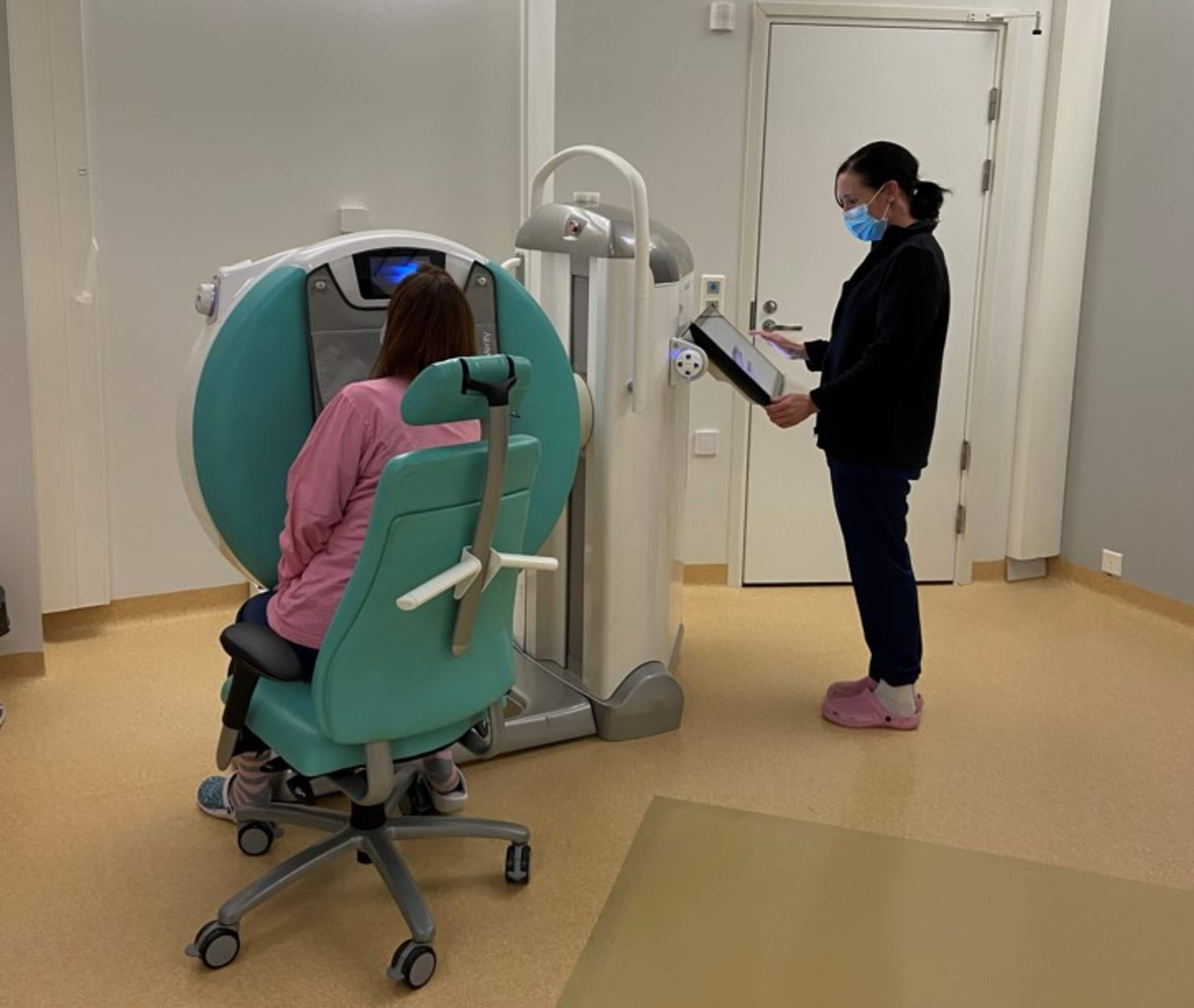
Cone beam CT examination is used for imaging areas such as the teeth or paranasal sinuses. The device can also be used to examine limbs, such as the hand, wrist, elbow, knee, ankle, or foot. Devices used for imaging the head and limbs differ from each other. 

The abbreviation CBCT is commonly used for this examination. It requires a specialized device that is not available in every radiology unit. 

The imaging uses X-ray radiation. The radiation dose from a CBCT examination corresponds at most to approximately two weeks of natural background radiation. During the imaging, the device captures images from multiple angles in a single session. It is important to remain still during the imaging to ensure image clarity. 

In head imaging, the device rotates around the head similarly to panoramic imaging of the teeth or jaws. Reasons for imaging the teeth and jaws include removal of wisdom teeth, placement of dental implants, and assessment of injuries or other changes in the teeth or jawbone. Reasons for imaging the paranasal sinuses include surgical planning or prolonged sinus infections. 

When imaging an limb, the rotating part inside the device moves, but this rotation is not noticeable during the imaging. Reasons for extremity imaging include suspected fractures, surgical planning, or monitoring the healing of a fractured bone. 

No special preparation is required for the examination. 

However, carefully review the patient instructions you have received. Reserve approximately one hour for the visit. 

During the imaging, you will either stand or sit. The position depends on the area being imaged. 

The examination lasts approximately 15–30 minutes. Before the imaging, the radiographer will inform you which clothes or jewelry need to be removed. The radiographer will assist you in finding the correct position for imaging. 

A contrast agent containing iodine may be injected into a joint using a thin needle. The need for contrast is determined by the referring physician or the radiologist. 

The examination does not require follow-up and does not impose restrictions on normal daily activities. 

If contrast agent is used, you will receive separate aftercare instructions. 

The radiologist will review the images and provide a report. The referring physician will inform you of the results and the next steps in your care. Contact your care unit if you do not have a scheduled appointment or call time. 

The risk from the examination is minimal, and the radiation dose to the imaged area is very low. 

Contrast agents may occasionally cause allergic reactions, though this is rare. Imaging staff will always ask about known allergies in advance. If an allergic reaction occurs, the imaging unit is equipped to manage it. 

Updated 23.10.2025