Go to page content

PET with computer tomography examination (PET-CT)

In a PET-CT examination, the human body is examined using isotopes and a computed tomography device i.e. CT.

PET-CT examination can be used to assess the spread of cancer. It is also utilized in the evaluation of inflammatory conditions and certain brain and heart diseases. The combination of PET and CT imaging allows for highly precise detection of pathological changes. 

The most common PET-CT examination is metabolic imaging using fluorodeoxyglucose (FDG). The tracer, which contains glucose i.e. sugar, accumulates in areas with increased metabolic activity, such as cancer cells. 

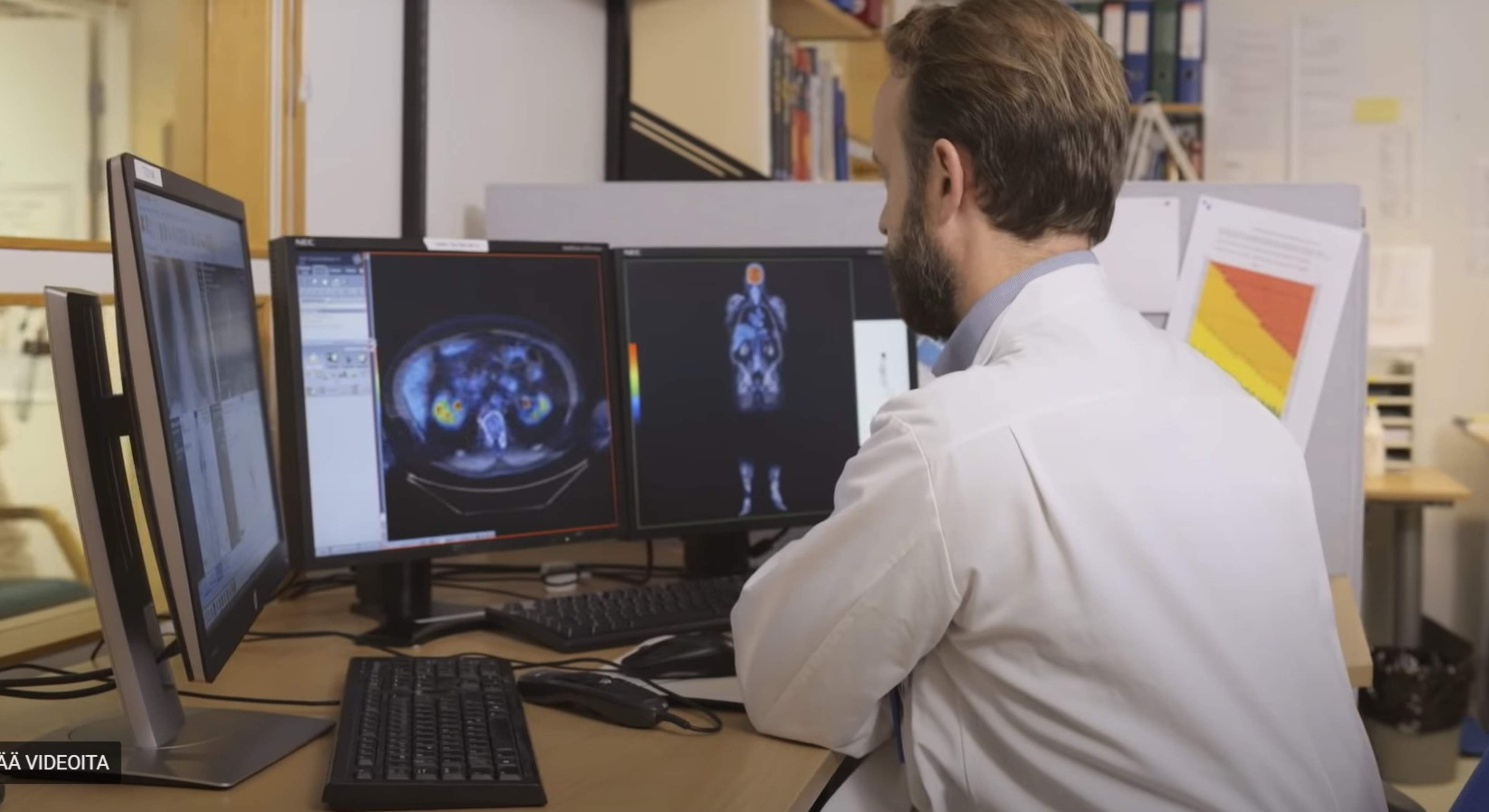
Watch the video: What is a PET examination? The video is produced by Turku PET Centre. Video is in Finnish with Swedish subtitles. 

Proper preparation is crucial for the success of the examination. Before the examination, you must refrain from eating and drinking and avoid physical exertion. For diabetic patients, blood glucose levels should be as close to normal as possible before the examination. You will receive detailed instructions in advance on how to prepare. 

If you know or suspect that you are pregnant, inform your treating physician or contact the imaging unit well in advanc

You will first be administered a radioactive tracer (radiopharmaceutical) intravenously. The radioactive tracer travels in the body to specific locations via metabolism or by binding to a receptor or a molecule on the cell surface. 

Before and after the tracer is administered, you will rest in bed. Resting helps to relax muscle tension and stabilize metabolism, allowing the glucose-containing tracer to better target the areas being examined. 

After resting, a low-dose CT imaging is performed to obtain a detailed structural image of the body. This is followed by a positron emission tomography (PET) imaging, which detects the locations where the tracer has accumulated in the body. Finally, the computer combines the CT and PET images to provide precise information about the location of any cancer or other disease in the body. 

The entire examination takes approximately 3 hours. 

After the examination, it is advisable to drink more fluids than usual and urinate frequently, as the radioactive tracer is excreted through urine. On the day of the examination, avoid close contact with pregnant individuals and refrain from holding children in your lap or sitting close to them. 

Breastfeeding mothers must take a break from breastfeeding after the scan. Milk expressed during this time should be discarded. The healthcare staff will provide detailed instructions to breastfeeding mothers after the examination. 

A radiologist will review and interpret the images and write a report. The physician who referred you for the examination will inform you of the results and discuss further treatment. Contact your care unit if you do not have a scheduled appointment or call time. 

The PET-CT examination is painless. 

The amount of radiation received from the imaging varies depending on the extent of the examination. The equipment used is highly sensitive and can detect very small amounts of radiation, meaning the risk from radiation exposure is low. Allergic reactions in isotope examinations are very rare. 

If you have concerns about radiation, you can always ask your treating physician or radiology staff for more information. You can also find information about radiation effects and radiation doses from different examinations in the Information about radiation section. 

Updated 28.11.2025