

Fundus photography is a quick and accurate way of diagnosing retinopathy, and it is the most common way of carrying out regular fundus screenings for diabetics. Fundus photography involves taking a photo of just the back of the eye, and it cannot be used to assess the health of any other parts of the eye.
An examination by an ophthalmologist is more extensive than fundus photography and involves a more comprehensive assessment of eye health. The ophthalmologist may check not only the structures of the eye but also how accurate your vision is and various eye pressure measurements. A fee is usually charged for an ophthalmologist's examination.
Sometimes, other examinations are required to carry out a more in-depth review of the retinas. These examinations can include optical coherence tomography (OCT) imaging and fluorescein angiography (injection of a coloured dye to check blood vessels and other structures).
How photographs are taken of the back of the eye
Photos of the back of the eye are taken with a fundus camera, and are then sent to an expert for review.
Eyedrops to dilate the pupils are used to help get the best possible photographs.
It only takes a few minutes to take the photographs, and it is not painful. It may cause you to be slightly dazzled.
After the photographs have been taken, your pupils may remain dilated for a few hours, during which time your eyes may be sensitive to light and your vision may be blurred.
You can prepare in advance by bringing sunglasses with you to your appointment.
It is advised that you do not drive for a couple of hours after having had the eyedrops.
Examination of the back of the eye by an ophthalmologist
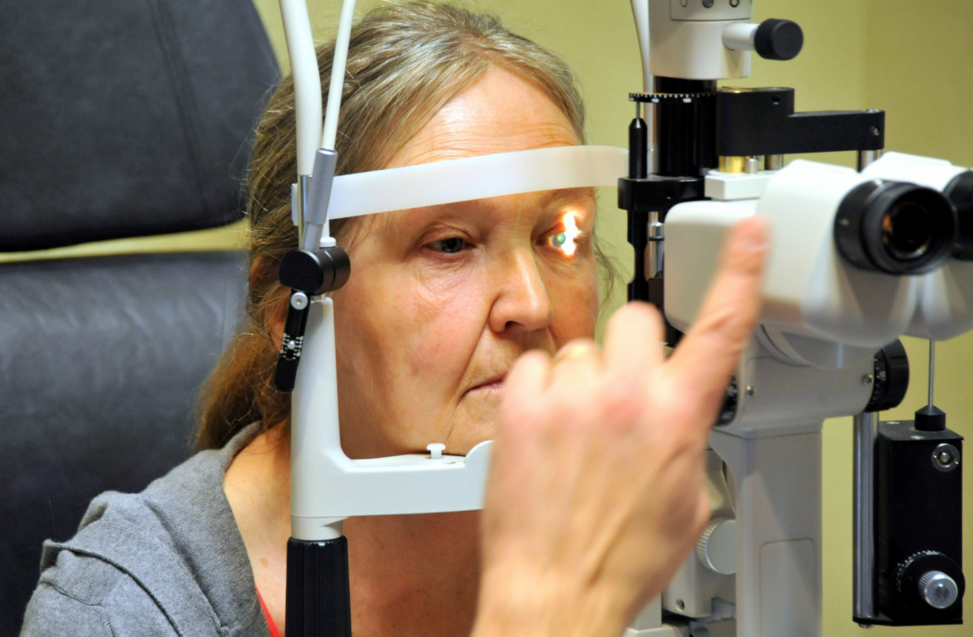
An ophthalmologist will examine the retina using an ophthalmoscope – an instrument used to examine the inside of the eye, or a biomicroscope. Preparations for this examination are the same as for fundus photography: pupil dilation eyedrops will be put in the eyes, you should prepare to be slightly dazzled, and driving is not advised for a couple of hours after having the eyedrops.